Ultrasound Template
Ultrasound Template - No evidence of tethering is seen. The sulcal/gyral pattern has a normal appearance for age and there is no midline shift. Click on the links below to open the worksheet as a downloadable pdf. The posterior bony structures are within normal limits with no evidence of dysraphism. Elbow pain, evaluate for tendon abnormality. Whilst not exhaustive it will remind users of the appearances of the more common lung pathologies. Web ultrasound, also called sonography, shows the structures inside the body. Included sections are the liver, gallbladder, bile ducts, pancreas, right kidney, left kidney, spleen, aorta, ivc, and abdominal fluid. An ultrasound picture is called a sonogram. No evidence of joint effusion or synovial process. Web structured radiology report template for a routine ultrasound of the abdomen. Web examples of reporting templates and anatomy. An ultrasound picture is called a sonogram. The cord terminates at t12 with a normal appearing conus. Web provide report templates for each individual ultrasound modality. Urinary tract and male reproductive system. No, it is not the conclusion. Web this article summarises the best practice in reporting of ultrasound examinations based on international literature and addresses key topics including report structure, clinical content, style and language. Elbow pain, evaluate for tendon abnormality. Web testicular and scrotal ultrasound is the primary modality for imaging most of the male reproductive system. Web structured radiology report template for a routine ultrasound of the abdomen. The right kidney measures __cm, the left kidney __cm. The endometrium is normal in appearance and thickness, measuring 5mm in double ap thickness. Web this article provides a beginners guide to ultrasound (pocus), including how ultrasound works and how ultrasound can be used in clinical practice. Web this report is designed as an educational aid aimed at guiding the user through the steps required to perform a lung ultrasound examination. Our templates are available in.rtf format, making them easily downloadable and compatible with powerscribe 360. Web structured radiology report template for ultrasound of the thyroid. Normal parenchymal echogenicity is seen. But most important the report tells much about you and your skills. No evidence of tethering is seen. The images can help guide diagnosis and treatment for many diseases and conditions. Web this article provides a beginners guide to ultrasound (pocus), including how ultrasound works and how ultrasound can be used in clinical practice. 1) your report is your business card. Browse our free ultrasound library offered to you by sonoskills and fujifilm healthcare europe. Click on the. Elbow pain, evaluate for tendon abnormality. Web learn how to scan plus how to interpret ultrasound images. The images can help guide diagnosis and treatment for many diseases and conditions. Comprehensive free guides describing equipment settings, probe placement, normal appearances, hints and tips. Web this article summarises the best practice in reporting of ultrasound examinations based on international literature and. Elbow pain, evaluate for tendon abnormality. Web this website is designed to host a set of radiology templates to use when reports are normal or near normal and to help standardize reports. Web learn how to scan plus how to interpret ultrasound images. No, it is not the conclusion. Web examples of reporting templates and anatomy. Web radreport.org provides reporting templates for many common radiology procedures that have been reviewed by an international panel of radiologists, as well as templates shared by members of rsna and the european society of radiology. Web this article summarises the best practice in reporting of ultrasound examinations based on international literature and addresses key topics including report structure, clinical content,. No evidence of joint effusion or synovial process. However, some involve placing a small device inside the body. The sulcal/gyral pattern has a normal appearance for age and there is no midline shift. Web ultrasound, also called sonography, shows the structures inside the body. Web structured radiology report template for a routine ultrasound of the abdomen. Web this report is designed as an educational aid aimed at guiding the user through the steps required to perform a lung ultrasound examination. Sample diagnostic elbow ultrasound report: So let us start with a few very important general facts and tips that will make you shine. No evidence of tethering is seen. Web radreport.org provides reporting templates for many. Web examples of reporting templates and anatomy. 1) your report is your business card. No evidence of joint effusion or synovial process. Numerous examples and sample phrases are provided and common pitfalls are discussed. Most ultrasounds are done using a device outside the body. It is relatively quick, relatively inexpensive, can be correlated quickly with the patient's signs and symptoms, and, most importantly, does not employ ionizing radiation. Structured templates for clear and consistent reports. The midline structures are unremarkable. Web this website is designed to host a set of radiology templates to use when reports are normal or near normal and to help. It is relatively quick, relatively inexpensive, can be correlated quickly with the patient's signs and symptoms, and, most importantly, does not employ ionizing radiation. Web testicular and scrotal ultrasound is the primary modality for imaging most of the male reproductive system. No evidence of joint effusion or synovial process. Urinary tract and male reproductive system. The sulcal/gyral pattern has a. No evidence of joint effusion or synovial process. Web structured radiology report template for a routine ultrasound of the abdomen. Web this article provides a beginners guide to ultrasound (pocus), including how ultrasound works and how ultrasound can be used in clinical practice. The right kidney measures __cm, the left kidney __cm. The cauda equina and filum terminalis are normal. An ultrasound picture is called a sonogram. The sulcal/gyral pattern has a normal appearance for age and there is no midline shift. The right kidney measures __cm, the left kidney __cm. The midline structures are unremarkable. Sample diagnostic elbow ultrasound report: Web 59552 ultrasound images & clips. Web learn how to scan plus how to interpret ultrasound images. Normal parenchymal echogenicity is seen. Normal intracranial anatomy is demonstrated. 1) your report is your business card. Elbow pain, evaluate for tendon abnormality. No, it is not the conclusion. The central echogenic complexes have a normal appearance with no hydronephrosis. Click on the links below to open the worksheet as a downloadable pdf. The endometrium is normal in appearance and thickness, measuring 5mm in double ap thickness. However, some involve placing a small device inside the body.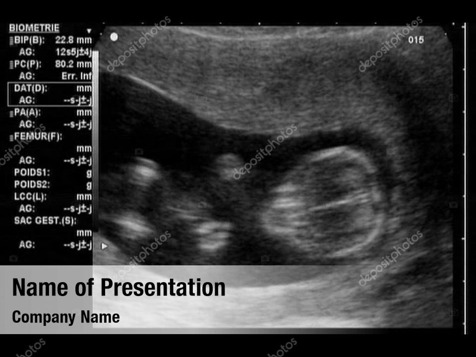
Ultrasound sonogram PowerPoint Template Ultrasound sonogram
Medical brochure or flyer template for ultrasound diagnostic, sonogram
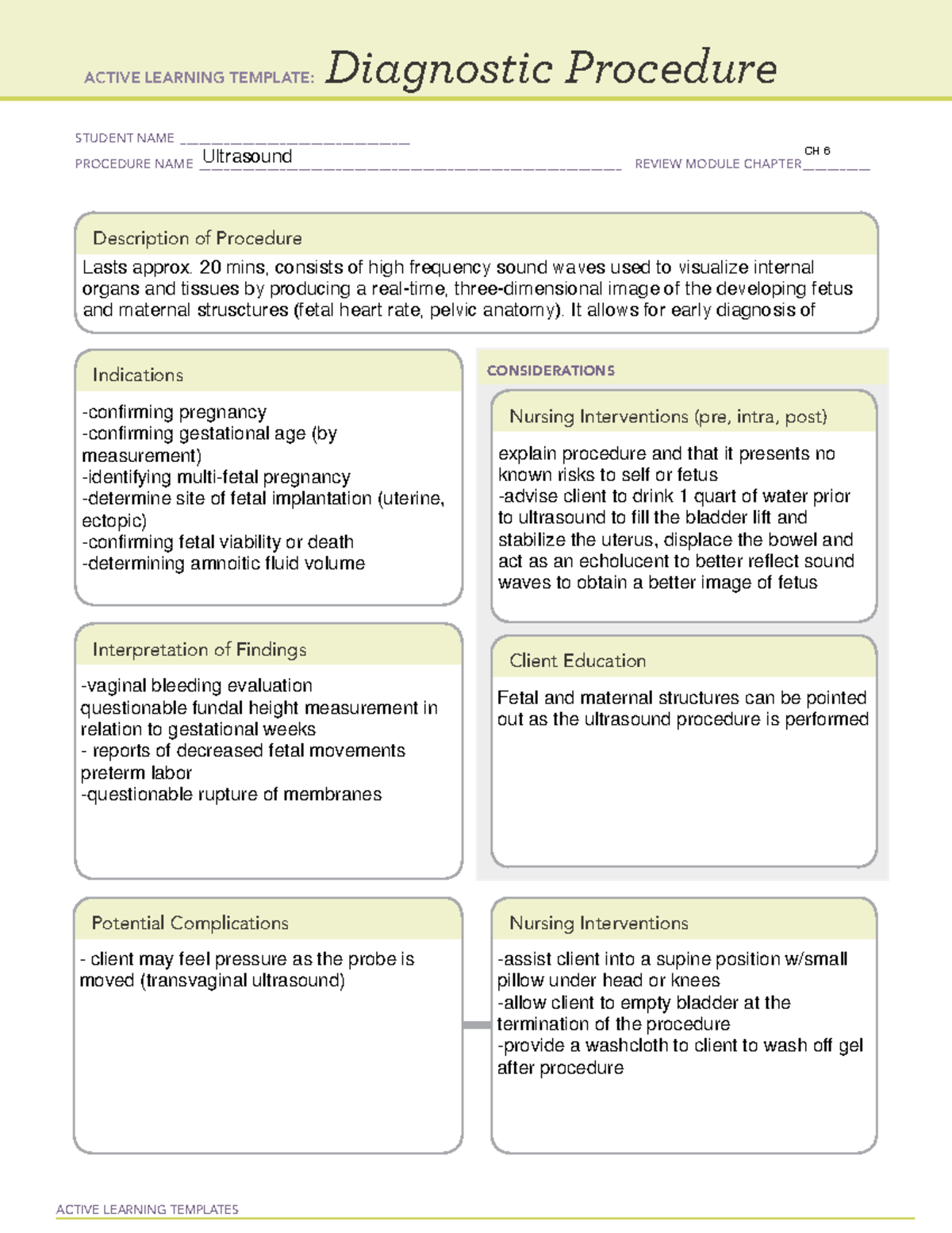
Ultrasound Diagnostic Procedure ATI Template.pdf ACTIVE LEARNING

The 10 Clinical Guidelines for the Perfect Ultrasound Report Format
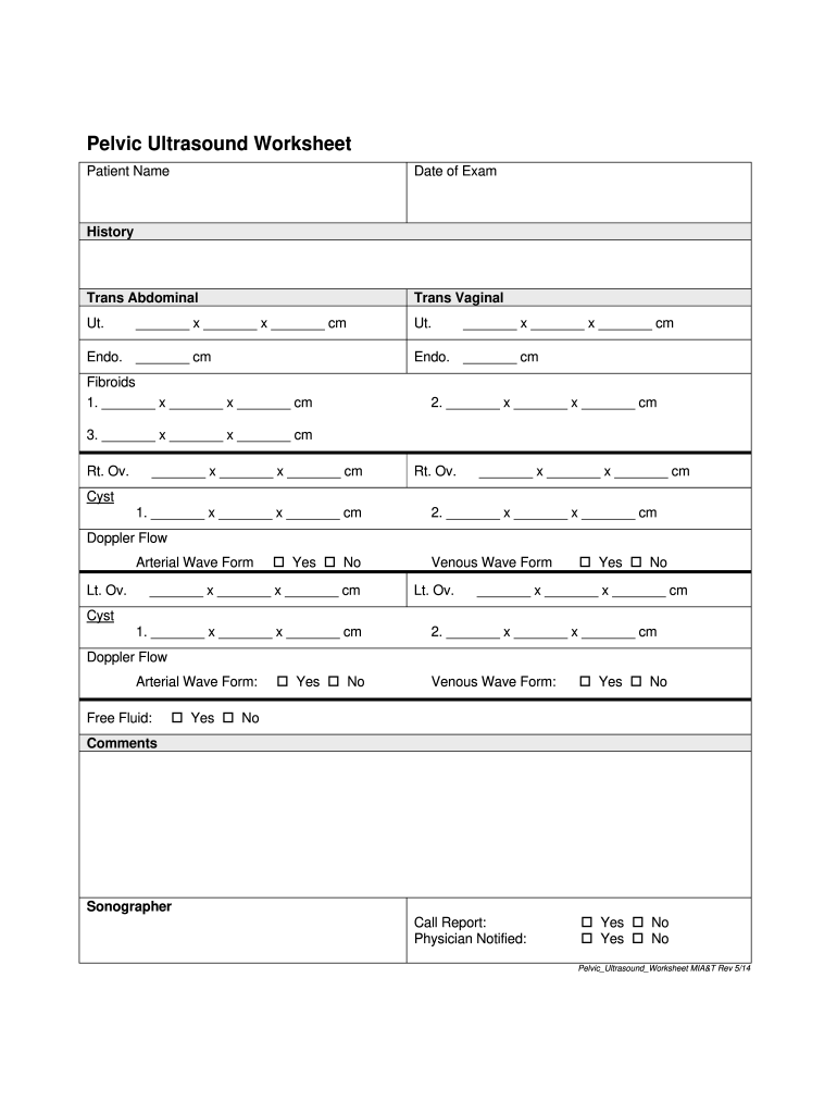
Free Printable Ultrasound Worksheets Free Printable Templates

Ultrasound template stock vector. Illustration of monitor 229779495

Printable Ultrasound Worksheets Fill Online, Printable, Fillable

Ultrasound scan of baby PowerPoint Template Ultrasound scan of baby

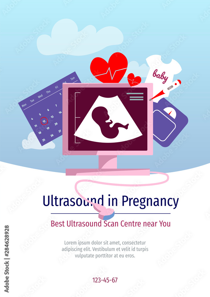
Premium Vector Ultrasound diagnostics flyer flat vector template
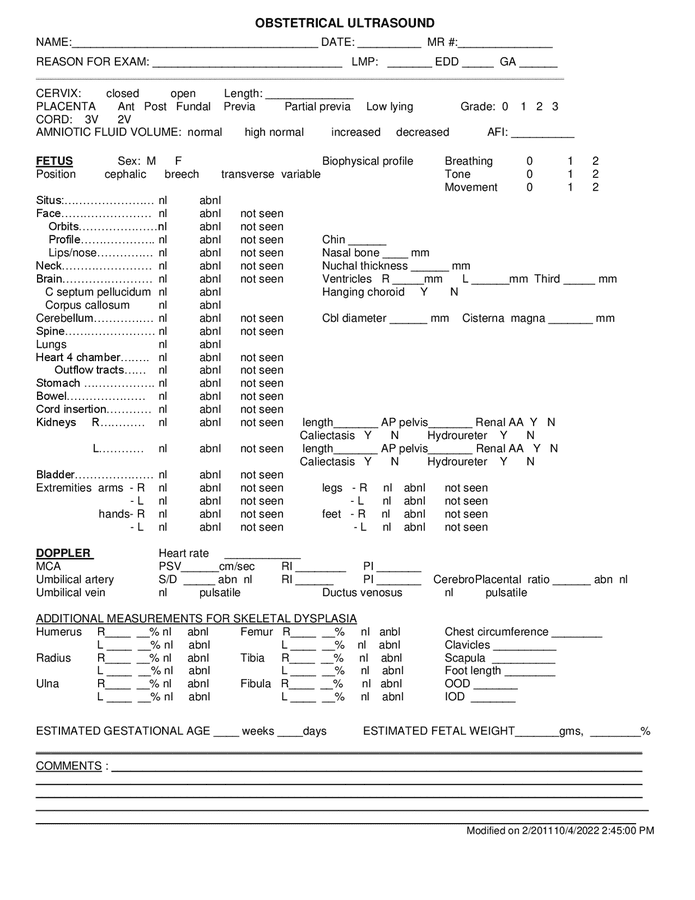
OB ultrasound report template in Word and Pdf formats
No Evidence Of Joint Effusion Or Synovial Process.
Whilst Not Exhaustive It Will Remind Users Of The Appearances Of The More Common Lung Pathologies.
The Images Can Help Guide Diagnosis And Treatment For Many Diseases And Conditions.
Web This Report Is Designed As An Educational Aid Aimed At Guiding The User Through The Steps Required To Perform A Lung Ultrasound Examination.
Related Post: